近日,2021届校友黄礼平、2009届校友孙洪伟所在研究团队成功将手持式拉曼光谱系统用于术中肝癌组织实时检测,探讨了利用拉曼光谱用于术中导航的可行性。此项研究成果于1月4日发表在国际顶级期刊《自然》(《Nature》)子刊。黄礼平校友为该论文第一作者。
拉曼光谱是一种分子振动光谱,基于振动分子的光非弹性散射可以提供复杂生物样本的化学指纹谱图。组织癌变进程中的生物成分变化,可以基于拉曼光谱以便捷、无损、无标记的形式得以反馈。但是由于光谱分析的复杂性以及癌组织的异质性,需要合适的数学分析模型辅助以实现来自不同组织类型的光谱数据识别与区分。对此,研究团队基于拉曼光谱和深度学习技术提出了一套用于肝癌组织体外和术中病理学诊断的工作流程。
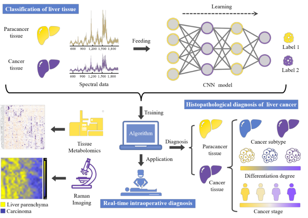
(图 基于拉曼光谱和智能算法的肝癌组织病理学诊断工作流程)
Abstract
Biopsy is the recommended standard for pathological diagnosis of liver carcinoma. However, this method usually requires sectioning and staining, and well-trained pathologists to interpret tissue images. Here, we utilize Raman spectroscopy to study human hepatic tissue samples, developing and validating a workflow for in vitro and intraoperative pathological diagnosis of liver cancer. We distinguish carcinoma tissues from adjacent non-tumour tissues in a rapid, non-disruptive, and label-free manner by using Raman spectroscopy combined with deep learning, which is validated by tissue metabolomics. This technique allows for detailed pathological identification of the cancer tissues, including subtype, differentiation grade, and tumour stage. 2D/3D Raman images of unprocessed human tissue slices with submicrometric resolution are also acquired based on visualization of molecular composition, which could assist in tumour boundary recognition and clinicopathologic diagnosis. Lastly, the potential for a portable handheld Raman system is illustrated during surgery for real-time intraoperative human liver cancer diagnosis.
2018年本科毕业于宁波大学海洋药学专业,2021年硕士毕业于温州医科大学生物医学工程专业,硕士期间研究课题为拉曼光谱医学诊断研究,导师为王毅研究员。2022年进入西湖大学纳米合成实验室继续攻读博士学位。
原文链接:
https://www.nature.com/articles/s41467-022-35696-2.

